Application notes

Technical notes

Ask an engineer


Publications
United States (EN)
Select your region or country.
PMTs for Microscopy Questions & Answers
- Why is high sensitivity better for confocal and multiphoton microscopy?
- What is a compound-semiconductor photocathode and how does it benefit confocal and multiphoton microscopy?
- What options are available to improve light collection for multiphoton imaging?
- How can I prevent damage to my detector during laser excitation?
Why is high sensitivity better for confocal and multiphoton microscopy?
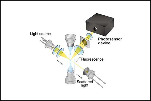
In confocal microscopy, a pinhole is used to remove extra light generated outside the focal point on the sample and to improve optical resolution and contrast. However, the amount of light that reaches the detector is reduced by the pinhole. So the detector needs higher sensitivity and lower dark current to achieve a better signal-to-noise ratio, resulting in higher dynamic range and in better contrast between the sample’s structure and the background.
Then how about multiphoton microscopy? Multiphoton microscopy does not require a pinhole like confocal microscopy, and it can observe deeper into the sample. Despite the light not being dimmed by a pinhole, the amount of signal is still very small because the multiphoton absorption process is a phenomenon that occurs with a very low probability. So a detector for multiphoton microscopy requires high sensitivity and low noise performance, just like confocal microscopy.
Hamamatsu offers both PMTs and MPPCs (SiPMs) as detectors in confocal and multiphoton microscopy, but the best detector depends on light intensity, sample thickness, scan speed, and many other factors. Please feel free to contact us regarding the selection of detectors.
What is a compound-semiconductor photocathode and how does it benefit confocal and multiphoton microscopy?
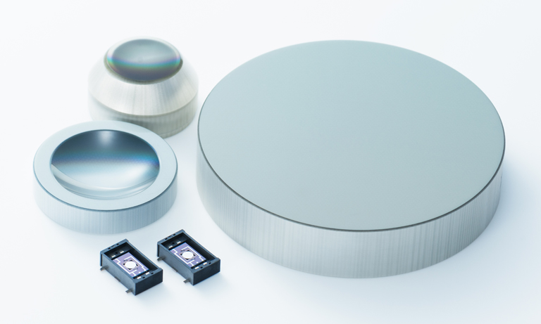
A compound-semiconductor photocathode for PMTs is designed to further improve the photocathode’s crystallinity and increase its sensitivity in the visible and near-infrared regions. To produce compound-semiconductor photocathodes, a crystal made of gallium arsenide (GaAs) or gallium arsenide phosphide (GaAsP) is adhered to glass, thinned to several µm thickness, and then reacted with cesium to achieve high quantum efficiency (QE). These compound-semiconductor photocathodes, which achieve high sensitivity, are superior to alkali photocathodes in the following ways:
- Since the crystalline film is thicker than other photocathodes, there is little light transmission loss, and the amount of light absorption and excited electrons generated is large.
- Because of its high crystallinity, the proportion of electrons and holes that recombine during the movement of electrons to the vacuum interface is small.
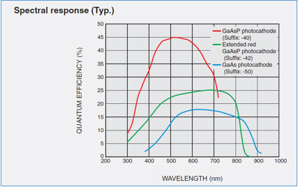
Figure 1. Typical spectral response curves for PMTs with GaAs and GaAsP photocathodes
As shown in the spectral response graph (Fig. 1), GaAs/GaAsP photocathodes are highly sensitive not only in the visible region but also in the red region (such as at 700 nm and 800 nm), which tends to have a smaller amount of fluorescence.
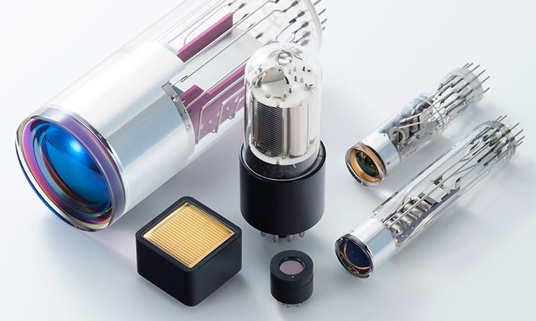
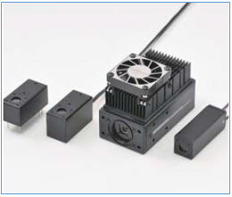
Although GaAs/GaAsP photocathodes have high QE, their disadvantage in the past was their low damage threshold. However, Hamamatsu has recently improved the durability of these photocathodes, and now offers several PMT modules (Fig. 2) that incorporate the improved photocathodes. Click on a part number below to learn more about these modules.
Among the modules listed above, the GaAsP PMT modules (-40 models) have improved maximum output current from 2 µA to 40 µA. The output current value of GaAs PMT modules (-50 models) has not been finalized, but it has improved.
In the H15460-40 GaAsP PMT module, the maximum current output has been further improved to 100 µA, the same value as a PMT with a typical alkali photocathode. The H15460-40 is discussed in more detail in the next question below.
What options are available to improve light collection for multiphoton imaging?
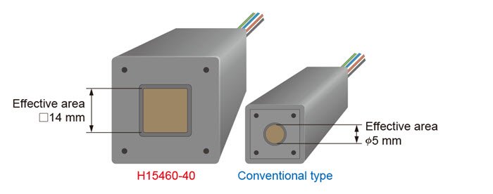
Figure 3. H15460-40 PMT module with a large 14 mm square effective area
To improve light collection in a multiphoton microscope, Hamamatsu offers the H15460-40 PMT module. This module’s 14 mm square effective area is larger than the 5 mm dia. of the PMT modules mentioned in the previous question (Fig. 3).
As described in a previous question, multiphoton microscopy obtains signals from a deeper part of the sample. The signals are diffused in the sample, and a detector with a wider field of view is required to collect as much of the signal as possible.
Also, when designing a microscope, it is difficult to balance the field of view, magnification, numerical aperture (NA), etc. For example, if you just want to widen the field of view, you can use a low-magnification objective lens, but this objective lens has a small NA. A detector with a large effective area helps facilitate such designs.
In addition to its larger area, the H15460-40 has a built-in amplifier to reduce noise between the detector and electronic devices, improving the overall sensitivity. This will improve the image contrast.
How can I prevent damage to my detector during laser excitation?
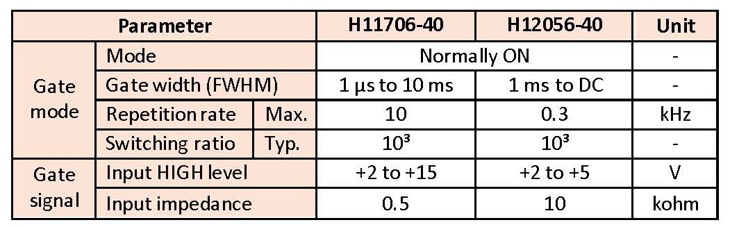
Table 1. Gating function specifications of H11706-40 and H12056-40 PMT modules
A gating function temporarily switches the distribution of the applied voltage to the dynode, preventing the electrons from moving to the subsequent stage. Please note that the gating function is effective only when the timing of the excess light is known in advance; it has no effect on accidental overexposure to light. See Table 1 for the gating specifications of the H11706-40 and H12056-40 PMT modules.
If you have a technical question you’d like to see answered on this page, email us.
Meet the engineer

Hiro Furuhashi is a marketing engineer in Hamamatsu’s San Jose, CA, office. He had been designing PMT modules for 9 years before moving to the US from Japan in 2019. He currently conducts market research on microscopy, in vitro diagnostics (IVD), and flow cytometry (FCM), and he also provides technical support as a PMT and PMT module specialist. He is a very active person, loves traveling, loves sports particularly surfing, and goes to the beach to surf almost every weekend.
- Confirmation
-
It looks like you're in the . If this is not your location, please select the correct region or country below.
You're headed to Hamamatsu Photonics website for US (English). If you want to view an other country's site, the optimized information will be provided by selecting options below.
In order to use this website comfortably, we use cookies. For cookie details please see our cookie policy.
- Cookie Policy
-
This website or its third-party tools use cookies, which are necessary to its functioning and required to achieve the purposes illustrated in this cookie policy. By closing the cookie warning banner, scrolling the page, clicking a link or continuing to browse otherwise, you agree to the use of cookies.
Hamamatsu uses cookies in order to enhance your experience on our website and ensure that our website functions.
You can visit this page at any time to learn more about cookies, get the most up to date information on how we use cookies and manage your cookie settings. We will not use cookies for any purpose other than the ones stated, but please note that we reserve the right to update our cookies.
1. What are cookies?
For modern websites to work according to visitor’s expectations, they need to collect certain basic information about visitors. To do this, a site will create small text files which are placed on visitor’s devices (computer or mobile) - these files are known as cookies when you access a website. Cookies are used in order to make websites function and work efficiently. Cookies are uniquely assigned to each visitor and can only be read by a web server in the domain that issued the cookie to the visitor. Cookies cannot be used to run programs or deliver viruses to a visitor’s device.
Cookies do various jobs which make the visitor’s experience of the internet much smoother and more interactive. For instance, cookies are used to remember the visitor’s preferences on sites they visit often, to remember language preference and to help navigate between pages more efficiently. Much, though not all, of the data collected is anonymous, though some of it is designed to detect browsing patterns and approximate geographical location to improve the visitor experience.
Certain type of cookies may require the data subject’s consent before storing them on the computer.
2. What are the different types of cookies?
This website uses two types of cookies:
- First party cookies. For our website, the first party cookies are controlled and maintained by Hamamatsu. No other parties have access to these cookies.
- Third party cookies. These cookies are implemented by organizations outside Hamamatsu. We do not have access to the data in these cookies, but we use these cookies to improve the overall website experience.
3. How do we use cookies?
This website uses cookies for following purposes:
- Certain cookies are necessary for our website to function. These are strictly necessary cookies and are required to enable website access, support navigation or provide relevant content. These cookies direct you to the correct region or country, and support security and ecommerce. Strictly necessary cookies also enforce your privacy preferences. Without these strictly necessary cookies, much of our website will not function.
- Analytics cookies are used to track website usage. This data enables us to improve our website usability, performance and website administration. In our analytics cookies, we do not store any personal identifying information.
- Functionality cookies. These are used to recognize you when you return to our website. This enables us to personalize our content for you, greet you by name and remember your preferences (for example, your choice of language or region).
- These cookies record your visit to our website, the pages you have visited and the links you have followed. We will use this information to make our website and the advertising displayed on it more relevant to your interests. We may also share this information with third parties for this purpose.
Cookies help us help you. Through the use of cookies, we learn what is important to our visitors and we develop and enhance website content and functionality to support your experience. Much of our website can be accessed if cookies are disabled, however certain website functions may not work. And, we believe your current and future visits will be enhanced if cookies are enabled.
4. Which cookies do we use?
There are two ways to manage cookie preferences.
- You can set your cookie preferences on your device or in your browser.
- You can set your cookie preferences at the website level.
If you don’t want to receive cookies, you can modify your browser so that it notifies you when cookies are sent to it or you can refuse cookies altogether. You can also delete cookies that have already been set.
If you wish to restrict or block web browser cookies which are set on your device then you can do this through your browser settings; the Help function within your browser should tell you how. Alternatively, you may wish to visit www.aboutcookies.org, which contains comprehensive information on how to do this on a wide variety of desktop browsers.
5. What are Internet tags and how do we use them with cookies?
Occasionally, we may use internet tags (also known as action tags, single-pixel GIFs, clear GIFs, invisible GIFs and 1-by-1 GIFs) at this site and may deploy these tags/cookies through a third-party advertising partner or a web analytical service partner which may be located and store the respective information (including your IP-address) in a foreign country. These tags/cookies are placed on both online advertisements that bring users to this site and on different pages of this site. We use this technology to measure the visitors' responses to our sites and the effectiveness of our advertising campaigns (including how many times a page is opened and which information is consulted) as well as to evaluate your use of this website. The third-party partner or the web analytical service partner may be able to collect data about visitors to our and other sites because of these internet tags/cookies, may compose reports regarding the website’s activity for us and may provide further services which are related to the use of the website and the internet. They may provide such information to other parties if there is a legal requirement that they do so, or if they hire the other parties to process information on their behalf.
If you would like more information about web tags and cookies associated with on-line advertising or to opt-out of third-party collection of this information, please visit the Network Advertising Initiative website http://www.networkadvertising.org.
6. Analytics and Advertisement Cookies
We use third-party cookies (such as Google Analytics) to track visitors on our website, to get reports about how visitors use the website and to inform, optimize and serve ads based on someone's past visits to our website.
You may opt-out of Google Analytics cookies by the websites provided by Google:
https://tools.google.com/dlpage/gaoptout?hl=en
As provided in this Privacy Policy (Article 5), you can learn more about opt-out cookies by the website provided by Network Advertising Initiative:
http://www.networkadvertising.org
We inform you that in such case you will not be able to wholly use all functions of our website.
Close